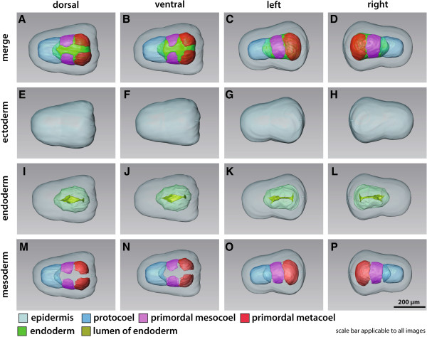
Figure 2.

3D reconstruction of the embryo of Saccoglossus kowalevskii at 36 h pf. Rows from left to right: dorsal, ventral, left, and right view. Columns from top to bottom: The merge row (A-D) shows the embryo with all reconstructed structures. Ectoderm row (E-H) shows external shape of embryo, telotroch not shown. Endoderm row (I-L) reveals the transparent endodermal tissue (light green) and the position of the endodermal lumen (yellowish green). Mesoderm row (M-P) shows the position of the anterior protocoel (blue) and the primordal meso- (pink) and metacoelic (red) tissue. Download interactive 3D-PDF as Additional file 1.
