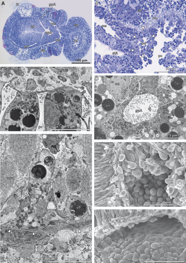
Figure 7.

Histology and fine structure of the late kink stage in Saccoglossus kowalevskii (~ 96 h pf). (A) sagittal section showing the internal organization. The anlagen of the 1st gill pores (gpA) are visible. The meso- (ms) and metacoelic (mt) cavities enlarge to form central lumina. (B) The pericardium (pd) is surrounded by a basement membrane (arrowheads) and furthermore bulb-shaped by protruding into the protocoelic cavity (pc). (C) The protocoelic cells lining the pericardium are differentiated into podocytes (po) resting on prominent blood sinus (bs) nested underneath the basement membrane (bm). (D) The elliptic anlage of the gill pore is constituted by monociliated cells though, the cytoplasm of individual cells contain several centrioles (ct) indicating ciliogenesis. (E) The majority of the protocoelic cells are differentiated into myoepithelial cells. Inner longitudinal muscle cells alternate with circular muscle cells as indicated by the arrangement of myofilaments (myo). (F) SEM of a dissected embryo revealing the goblet-shape of the apical cell processes of the protocoelic cells. (G) The meso- and metacoelic lining cells constitute a sqamous epithelium holding a single cilium (ci) at the cell surface. dms distal mesodermal cell, ed endoderm, ep epidermis, gpc gill pore cell, mi mitochondrion, nn nerve net, nu nucleus, pdc pericardial cell, pe pedicels, pms proximal mesodermal cell, pph primordal pharynx, stA anlage of the stomochord, yo yolk, za zonula adherens.
