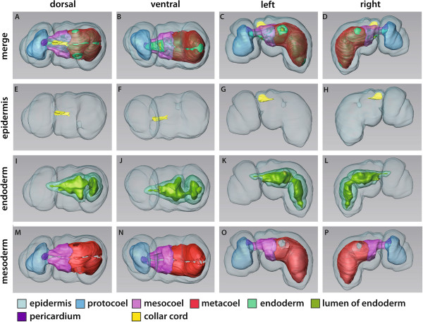
Figure 8.

3D-reconstruction of the embryo of Saccoglossus kowalevskii at 1 gill slit stage (~ 132 h pf). Rows from left to right: dorsal, ventral, left and right view. Columns from up to down: The merge row (A-D) shows the embryo with all reconstructed structures. The first gill pore (gp) is opened on the left side whereas the right side is still closed. Epidermis row (E-H) shows the external shape of embryo. The telotroch is not shown. The collar cord (cc) is invaginated by a process similar to chordate neurulation. Endoderm row (I-L) shows the transparent endodermal tissue (light green) revealing the right-curved course of the oesophagus (oe). Only the left gill pore is opened. The still short stomochord (st) is protruding into the protocoel. At the posterior end of the digestive tract a hindgut region (hg) can be distinguished. Mesoderm row (M-P) shows the position of the anterior protocoel (blue) and the paired meso- (pink) and metacoelic (red) compartments. The pericardium (purple) is protruding into the protocoelic cavity. Download interactive 3D-PDF as Additional file 6. i intestine, mo mouth opening, ph pharynx.
