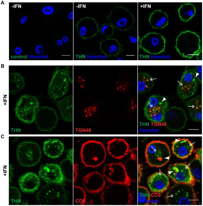
Figure 2. Tetherin localises to the cell surface, TGN, and IPMCs.
(A) Untreated MDMs (−IFN), or MDMs treated for 24 h with 544 U/ml IFN-β (+IFN), were incubated for 1 h on ice in media containing 10 µg/ml polyclonal Tetherin (THN) antibody (B02P), or VSV-G antibody as a negative control. Cells were fixed and labelled with a fluorescent secondary antibody. (B–C) IFN-β-treated MDMs were incubated for 20 min on ice with 10 µg/ml polyclonal Tetherin antibody (B02P) and 2.5 µg/ml anti-TGN46, or with 10 µg/ml monoclonal Tetherin antibody (M15) and 2 µg/ml anti-CD9, in the presence of 0.05% saponin. Cells were fixed and labelled with fluorescent secondary antibodies. Arrowheads point at structures reminiscent of IPMCs, double arrows indicate TGN-like staining patterns. All images are single confocal sections. Scale bars = 10 µm.

