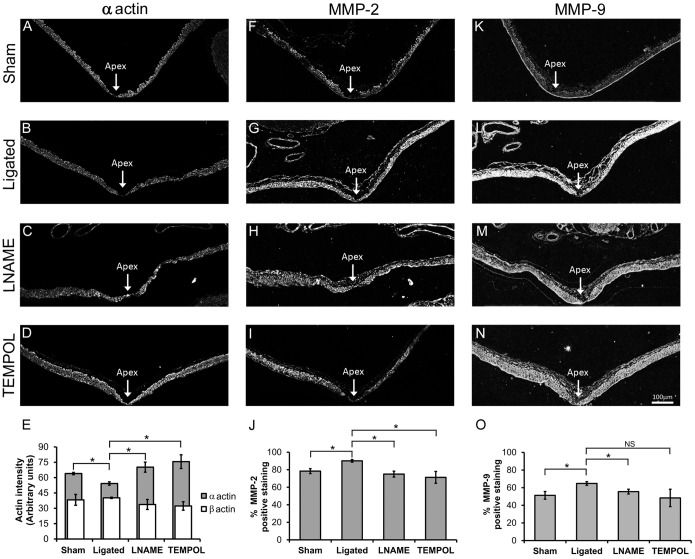
Figure 7. LNAME and TEMPOL restored α-actin and decreased MMPs at the BT of ligated animals.
Smooth muscle α-actin, MMP-2 and MMP-9 proteins were detected by immunofluorescence in sham-operated animals (A, F, K), and in CCA ligated animals given no inhibitors (B, G, L), or treated with LNAME (C, H, M), or TEMPOL (D, I, N). α-actin staining in the media significantly decreased at the BT following ligation (B) compared to sham-operated animals (A), but was restored with either LNAME (C) or TEMPOL (D) treatment. MMP-2 staining increased at the BT following ligation (G) compared to the sham surgery group (F), and was decreased with either LNAME (H) or TEMPOL (I) treatment. Similarly, MMP-9 staining in the media increased at the BT following ligation (L) compared to sham-operated animals (K), and was decreased with either LNAME (M) or TEMPOL (N) treatment. Scale bar = 100 µm. Panels E, J and O show quantitative analysis of these stainings. (E) Intensity of α-actin and β-actin were measured in SMCs at the BT. α-actin was decreased by ligation but increased with both LNAME and TEMPOL treatments while β-actin did not change among groups. Quantification of MMP-2 (J) and MMP-9 (O) immunofluorescence indicated significant changes in the percent area of positive staining at the BT. While TEMPOL treatment decreased MMP-9 staining compared to untreated ligated animals the change was not statistically significant. Bars represent mean ± standard error. *Statistically significant difference between groups (p≤0.05, Mann-Whitney U-test). NS indicates no significant difference.

