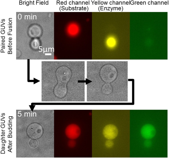
Figure 2. Fusion, reaction, and budding of vesicles.
The upper images show GUVs before fusion. The red channel shows the marker for the substrate-containing GUV, whereas the yellow channel shows the marker for the enzyme-containing GUV. The middle images show the budding transformation process after electrofusion. The lower images show daughter GUVs after budding. The increased fluorescence in the green channel indicates the occurrence of the enzymatic reaction.

