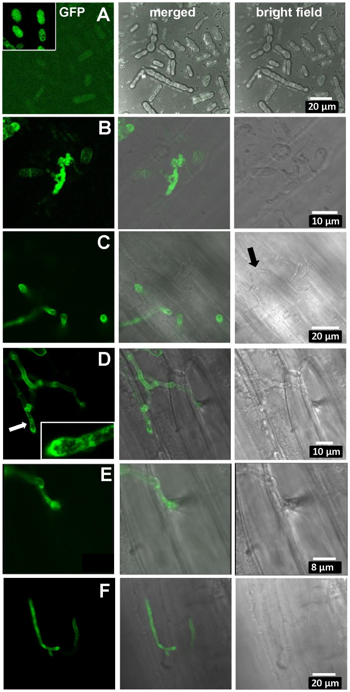
Figure 6. Expression of UhAVR1:GFP chimers during infection.
Confocal microscopy of mated U. hordei strains transformed with various GFP constructs. A. Free-floating and mated cells of cross Uh1353×Uh362 (MAT-2, Uhavr1) showing no green fluorescence, whereas GFP expressed from the strong constitutive otef promoter in strain Uh364 (Uh1351) shows bright fluorescence (insert); protein blot analysis verified no expression from Uh1353 and strong expression of just GFP from Uh1351 under these conditions (Figure S9B). B. As a control, Uh1357 (MAT-1 ΔUhAvr1 [otef:UhAvr1:GFP])×Uh362 on compatible Odessa coleoptiles at 48 hai shows strong GFP expression from the otef promoter in the same recipient strain. C. Uh1353×Uh362 on compatible Odessa coleoptiles at 48 hai shows septated hyphae on the surface devoid of cytoplasm and not fluorescing (arrow) whereas in invaded dikaryotic hyphae expression of the UhAVR1:GFP chimer is induced from its native promoter upon host “sensing” and penetration. D. As in C but at 100 hai, showing extended, septated hyphae. Fluorescence is visible in the hyphal cell wall but appears punctuated seemingly in vesicle-like structures in the growing points being concentrated at the tip (insert). E. Enlargement from D of penetration site on the right. F. Same cross as in C induces UhAvr1 expression in incompatible Hannchen at 48 hai, but no HR is seen.

