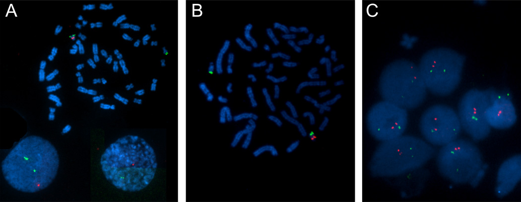
Figure 2. Results of fluorescence in situ hybridization analysis of cultured peripheral-blood lymphocytes from the VCFS/DGS son and his mother.
A representative metaphasic spread of chromosomes are shown for the Son (2A) and Mother (2B). The green dots are chromosome 22 control probes (ARSA; 22q13) and the red dots are the 22q11.2 probes (TUPLE1). The son (2A) appears to have one signal for the 22q11.2 probe, showing a deletion of the 22q11.2 region. The mother’s chromosomes during metaphase (2B) have one signal for the 22q11.2 probe (red). However when the mother’s chromosomes were analyzed during interphase (2C), it is evident that two signals for the 22q11.2 probe are seen, likely resulting from a duplication of the 22q11.2 region. This was not seen in the interphase analysis of the son (Figure 2A).

