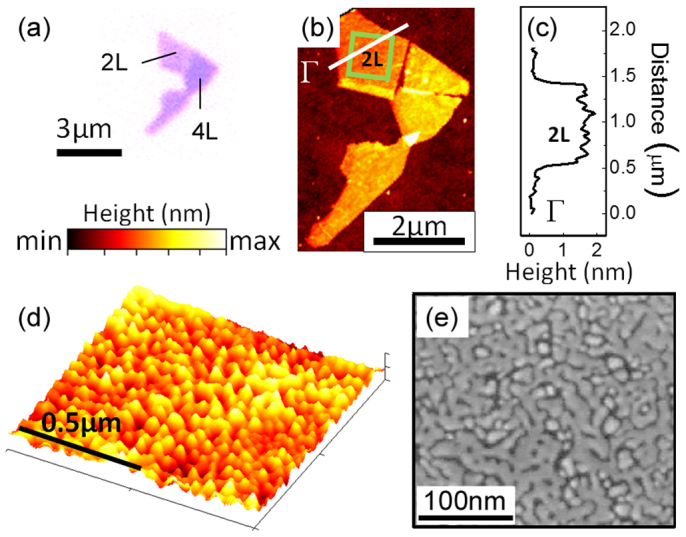
Figure 1. Exfoliated MoS2 flake and 2.0 nm Au deposition.

(a) Optical image of exfoliated MoS2 flake. Different colors correspond to the different number of MoS2 layers: 2 layers (2 L) and 4 layers (4 L). (b) AFM image of the MoS2 flake as exfoliated (color scale: min = 10.4 nm, max = 18.4 nm). Γ line (black) corresponds to the profile cross-section extracted (c) to confirm the height of the two layer. (d) High resolution AFM image Au nanoislands on MoS2 corresponding to boxed area (green) in (b) (color scale: min = 0.0 nm, max = 2.9 nm) (e) SEM image of Au nanostructures on MoS2.
