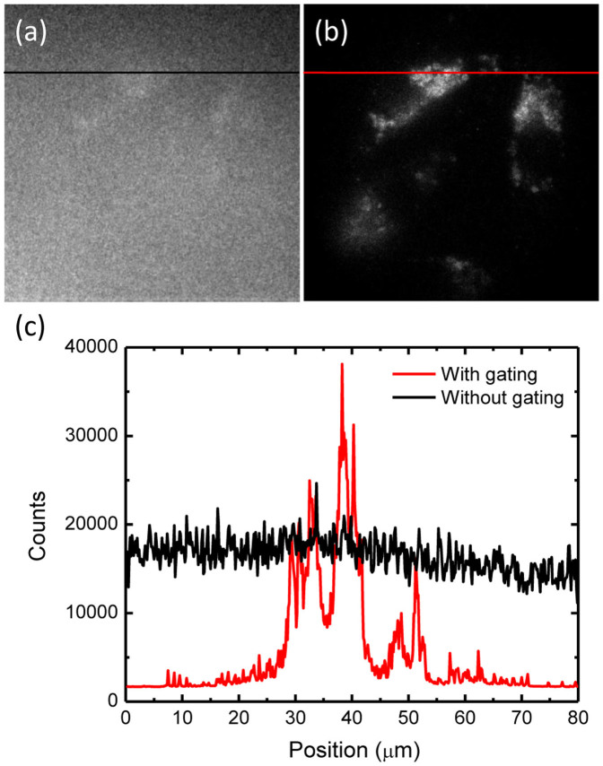
Figure 3. In vitro imaging of FND-labeled cells in blood.
(a, b) Wide-field fluorescence images of FND-labeled HeLa cells attached to a coverglass slide and immersed in human blood without (a) or with (b) time gating. The exposure times used for the fluorescence imaging with a 100× oil-immersion objective lens in (a) and (b) are 0.1 s and 0.3 s, respectively. (c) Intensity profiles along the black and red color lines denoted in (a) and (b), respectively.

