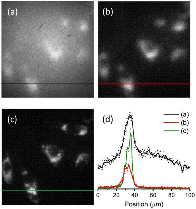
Figure 4. In vitro imaging of FND-labeled cells covered with chicken breast.
(a, b) Wide-field fluorescence images of FND-labeled HeLa cells in blood on a glass slide covered with a thin layer (~0.1 mm thickness) of chicken breast (a) without and (b) with time gating. (c) Time-gated fluorescence images of the corresponding FND-labeled HeLa cells in (a) and (b) without chicken breast. The objective lens is 40×. (d) Intensity profiles along the black, red color, and green lines denoted in (a), (b), and (c), respectively.

