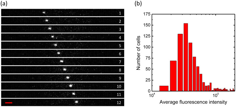
Figure 5. Flow cytometric analysis of FND-labeled cells in a microchannel.
(a) Snapshots of the flow of a single HeLa cell labeled with 100-nm FNDs in a microchannel with human blood. The channel width is 50 μm, the frame rate is 23 Hz (i.e. 43.5 ms/frame), and the objective lens is 10×. The red scale bar corresponds to 50 μm. (b) Histogram of the FND-labeled cells flowing across the microchannel with a cell concentration of 1.7 × 10⁵ cells/mL and a pumping rate of 0.01 mL/h for 2155 s. The expected and counted numbers of the cells are 998 and 862, respectively.

