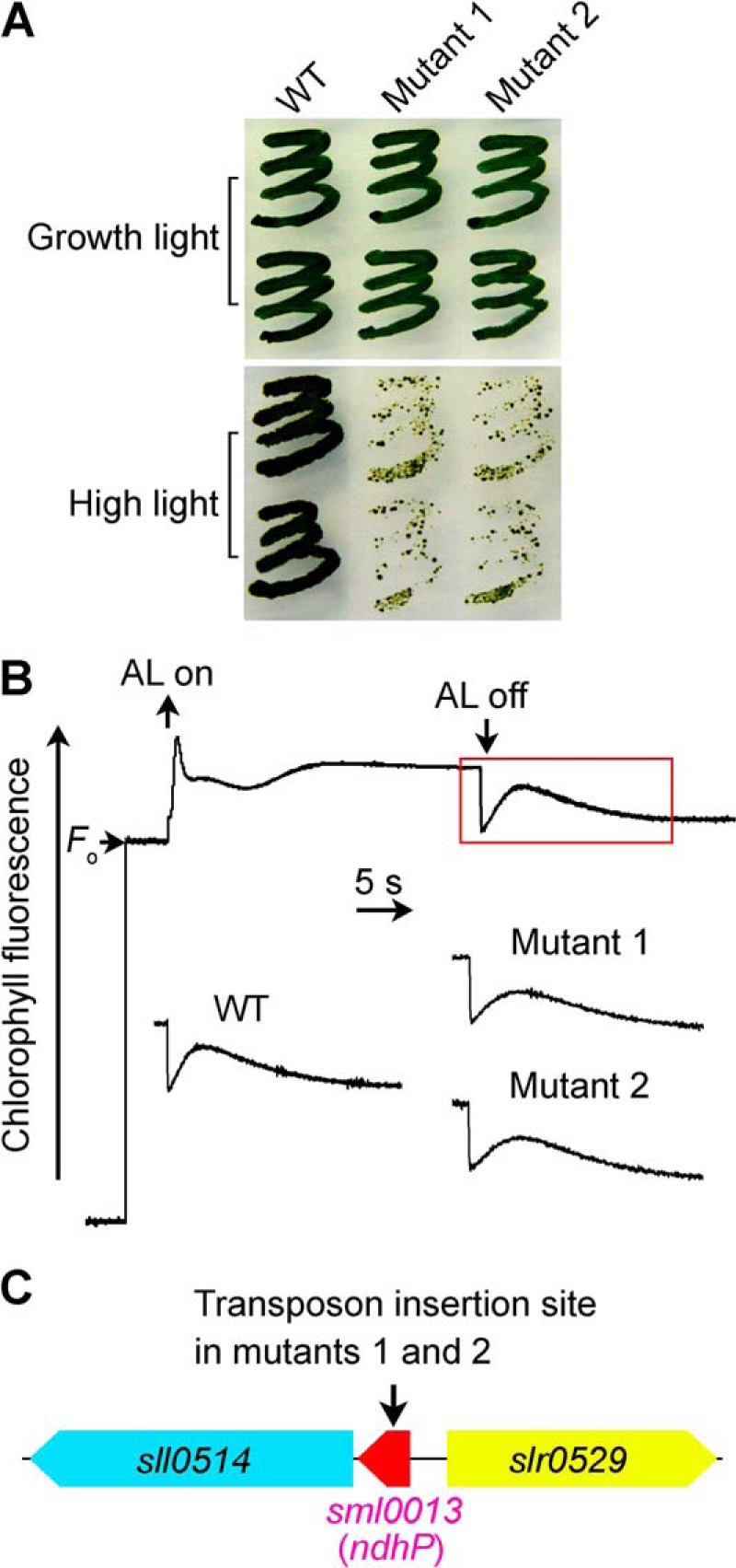
FIGURE 1.

Growth, NDH-CET activity, and transposon insertion site of high light-sensitive mutants of Synechocystis 6803. A, growth of WT and 2 mutants under normal light (40 μE m−2 s−1) and high light (200 μE m−2 s−1). B, monitoring of NDH-CET activity using chlorophyll fluorescence analysis. The top curve shows a typical trace of chlorophyll fluorescence in the WT Synechocystis 6803. See “Experimental Procedures” for experimental details. C, arrow schematically indicates the transposon insertion site in mutants 1 and 2 probed by PCR analysis using the primers listed in supplemental Table 1.
