
FIGURE 3.
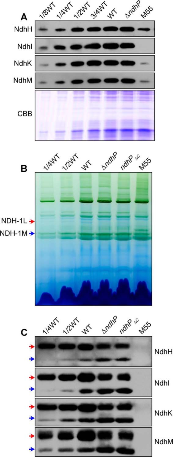
Western analyses of NDH-1L and NDH-1M complexes from the WT, ΔndhP, ndhPΔC, and M55 strains. A, Coomassie Brilliant Blue (CBB)-staining PAGE profiles and Western analysis of total NDH-1 in the thylakoid membranes. B, profiles of BN-PAGE of the thylakoid membranes; C, Western analysis of NDH-1L and NDH-1M complexes (indicated by red and blue arrows, respectively) from the WT, ΔndhP, ndhPΔC, and M55 strains. Sample containing 9 μg of chlorophyll a was loaded onto each lane.