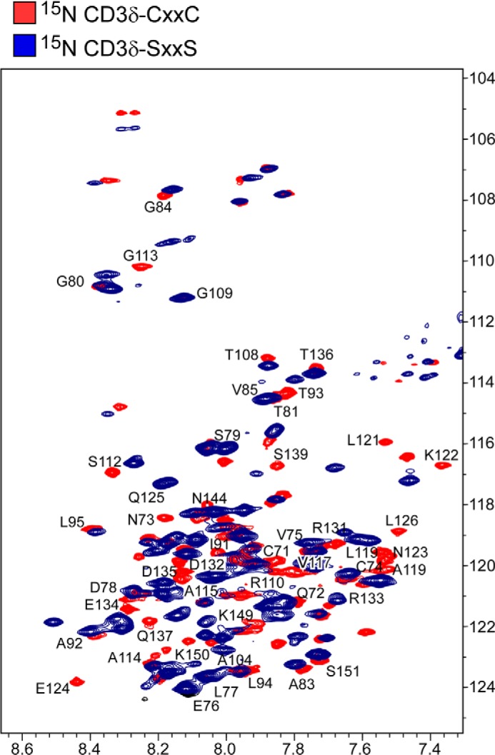
FIGURE 6.

Structural alterations in CD3δ by SXXS mutation. 1H,15N TROSY HSQC spectrum of CD3δ-CXXC (blue) is overlaid with the TROSY HSQC spectrum of CD3δ-SXXS (red). Representative resonances are labeled in the membrane proximal, transmembrane, and cytoplasmic tail of CD3δ. Numbering corresponds to the full-length murine wild type CD3δ protein where the CXXC motif is at residues 71–74 (Fig. 1A).
