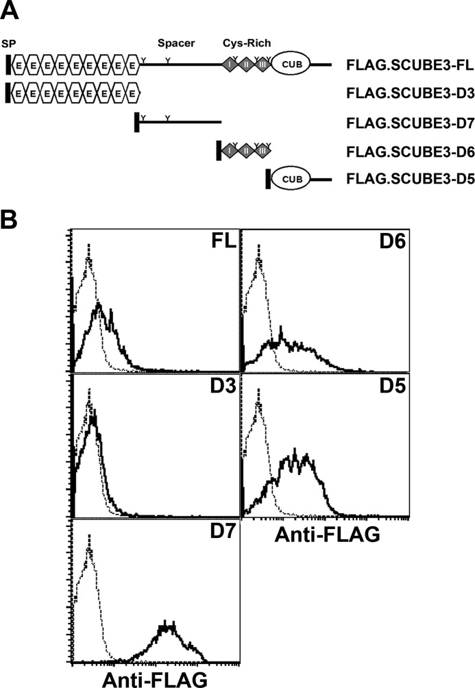
FIGURE 5.
Flow cytometry of cell surface expression of SCUBE3. A, domain structure of SCUBE3-FL and its deletion constructs. A FLAG tag was added immediately after the signal peptide sequences, thus at the NH2 terminus, for easy detection. FL, amino acids 1–993; D3, amino acids 1–401; D6, amino acid 398–629; D7, amino acids 632–803; D5, amino acids 804–993. FL, full-length; SP, signal peptide; E, EGF-like repeats; Cys-Rich, cysteine-rich motifs; CUB, CUB domain. B, the spacer region, cysteine-rich motifs, or CUB domain of SCUBE3 is capable of tethering on cell surface. At 24 h after transfection of empty vector or these expression plasmids, cells were detached and stained with anti-FLAG monoclonal antibody to determine the cell surface expression by flow cytometry. Empty vector-transfected cells are presented as the basal expression level (dotted line), and SCUBE3-transfected cells are shown as a thick line.

