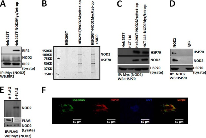
FIGURE 2.
Interaction between NOD2 and HSP70. A, co-IP (IP) were performed using 1 μl of mouse anti-Myc antibody per 150 μl of lysate (HEK293T-NOD2Myc/Tet-op, stimulated with 2 μm of MDP) and probed for RIP2 using rabbit anti-RIP2 antibody (1:1000). WB, Western blot. B, SYPRO®-stained 7.5% polyacrylamide gels showing the bands that were obtained through co-immunoprecipitation using mouse anti-Myc antibody in HEK293T, HEK293T-NOD2Myc/Tet-op (no dox), and HEK293T-NOD2Myc/Tet-op (no dox) cells stimulated with 2 μm of MDP. The visible bands were characterized using protein mass spectrometry as described under the “Experimental Procedures.” NOD2 and HSP70 were identified. C, co-IP performed using mouse anti-Myc antibody (1 μl/150 μl of lysate) in HEK293T-NOD2Myc/Tet-op (no dox), HCT 116-NOD2Myc/Tet-op (no dox), HEK293T, and HCT 116 cells and probed for HSP70 using rat anti-HSP70 antibody (1:1000) and rabbit anti-Myc antibody (1:1000). D, co-IP performed using rabbit NOD2 antiserum HM2559 (2 μl/150 μl) or rabbit IgG in THP-1 cells and probed for HSP70 using rat anti-HSP70 antibody (1:1000) and NOD2 using rabbit NOD2 antiserum HM2559 (1:5000). E, co-IP performed using mouse anti-FLAG antibody (5 μl/150 μl of lysate) in HEK293T cells transfected with pBKCMV/ATPase (A-FLAG) or pBKCMV/substrate binding domain (S-FLAG). Western blot was performed to detect NOD2 using Myc antibody (1:1000) and FLAG using FLAG antibody (1:500). F, HEK293T cells transiently transfected using pBKCMV/NOD2Myc vector and stained for NOD2 using mouse anti-Myc antibody (1:2000) and rabbit anti-HSP70. DAPI was used to stain the nucleus. WB, Western blot.

