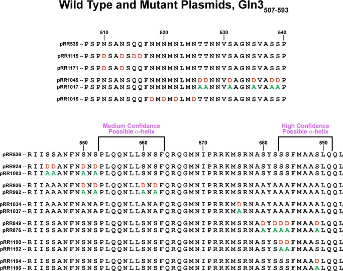
FIGURE 1.
Gln3 protein sequences analyzed in this work. The wild type Gln3 sequence (pRR536) appears at the top along with coordinates, relative to the first methionine. Single or paired sequences below the wild type are those of the mutant plasmids; plasmid numbers appear beside the mutant sequence. Aspartate and alanine substitutions are indicated in red and green, respectively. All mutant plasmids contain a full-length gln3 gene driven by its native promoter. Two sequences predicted as potentially folding into α-helices are indicated.

