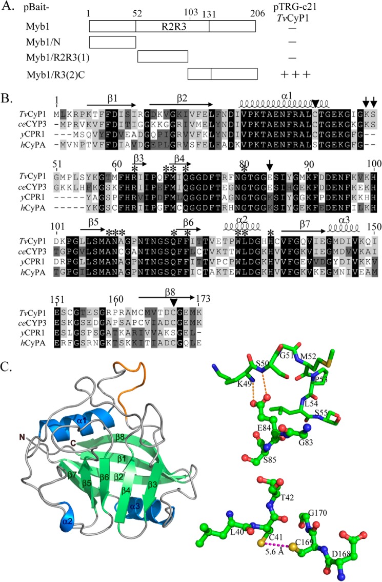
FIGURE 1.
Interaction of TvCyP1 with various regions of Myb1 and sequence analysis of TvCyP1. In A, DNA fragments from various regions of the myb1 gene were cloned into pBait to react with TvCyP1-expressing pTRG-c21 in a bacterial two-hybrid assay. The relative strength of the interaction as revealed by the formation of colonies in each assay (−, no colony formation; +++, >300 colonies) is summarized in the right panel. In B, the sequence of TvCyP1 was aligned to that of CeCyP3 (P52011), yCPR1 (P14832), and hCyPA (P62937). The amino acids involved in the enzyme activity and CsA binding are indicated by asterisks. The amino acids involved in hydrogen bond formation are indicated by arrows, and those in disulfide bond formation by arrowheads. In C, the predicted three-dimensional structure of TvCyP1 using CeCyP3 as a template is depicted in the left panel. Local aa sequences in TvCyP1 are respectively depicted in ball-and-stick modes. The dotted lines represent hydrogen bond formation between Glu84 and Lys49/Ser50 or disulfide bond formation between Cys41 and Cys169.

