FIGURE 8.
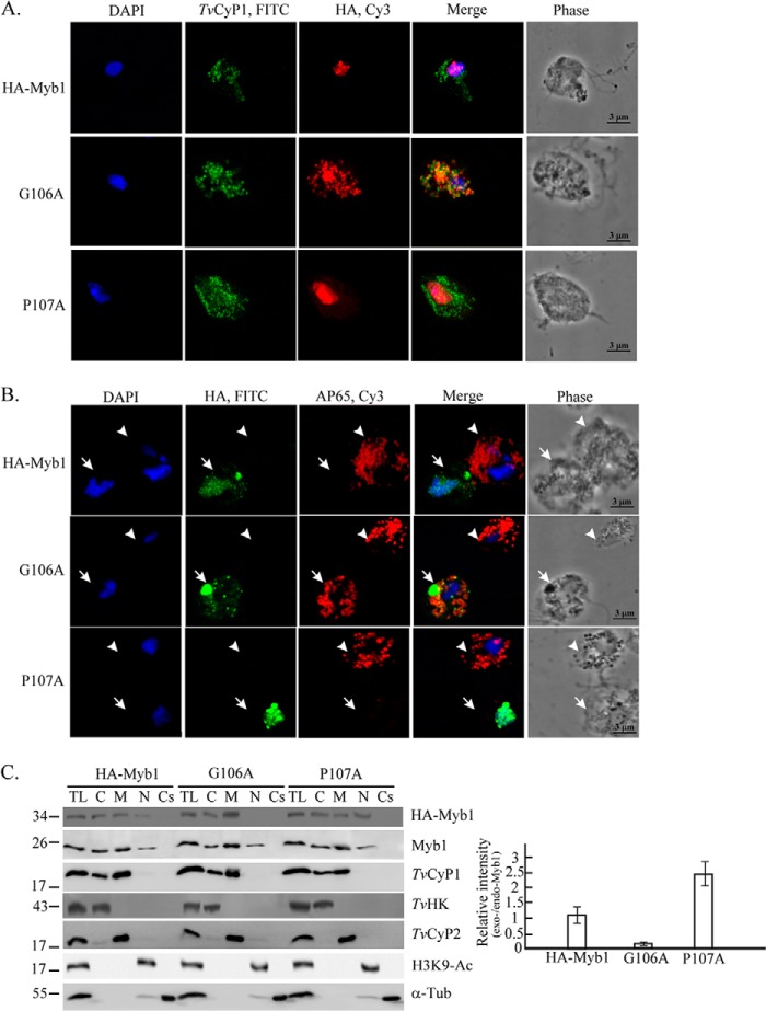
The role of TvCyP1-binding motif in the trafficking of Myb1 to the nucleus. Transfected cells overexpressing HA-Myb1, HA-Myb1(G106A), and HA-Myb1(P107A) were examined by IFA (A and B). Slides were double stained, first with either rat anti-TvCyP1 (A) or rat anti-HA (B) followed by FITC-conjugated rat IgG and then with either mouse anti-HA (A) or 12G4 antibody (B) followed by Cy3-conjugated mouse IgG as indicated at the top. Nuclei were stained with DAPI. Fluorescence signals were each recorded under confocal microscopy and merged. Transfected and nontransfected cells in B are indicated by open arrows and open arrowheads, respectively. Cell morphology was recorded under phase-contrast microscopy. The bar in the micrographs represents 3 μm. In C, total cell lysates (TL) from transfected cells and controls were fractionated into cytosolic (C), membranous (M), nuclear (N), and cytoskeletal (Cs) fractions for Western blotting using rat anti-HA and mouse anti-Myb1 antibodies to detect overexpressed (exo-Myb1) and endogenous Myb1 (endo-Myb1), respectively. Blots were also probed using the antibodies to detect various proteins as indicated. The relative intensities of overexpressed Myb1 versus endogenous Myb1 in nuclear samples from three separate experiments are quantified in histograms. Error bars represent S.D. from three separate experiments. Tub, tubulin.

