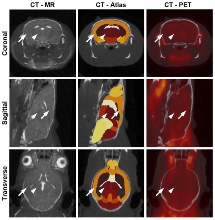
Fig. (1).
Trimodality mouse imaging with CT, PET and MR. An example of a 5XFAD mouse with overlay of CT with MR (left) demonstrates brain registration between these modalities. Parcellation of the brain into various regions was accomplished with MR data and aligned with the CT (middle). 18FDG-PET aligned with the CT (right) was evaluated for whole and regional brain uptake. The neocortex (arrow) and thalamus (arrowhead) are identified as examples of parcellated brain regions.

