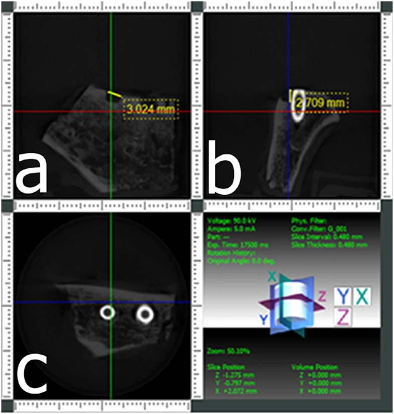
Figure 3.
Cone beam CT images from a scan obtained at a 40 × 40 mm field of view of an implant with a buccal marginal alveolar peri-implant defect shown in Figures 1, 2. (a) A coronal section providing the width of the defect, (b) a cross-sectional section providing the depth of the defect and (c) an axial section.

