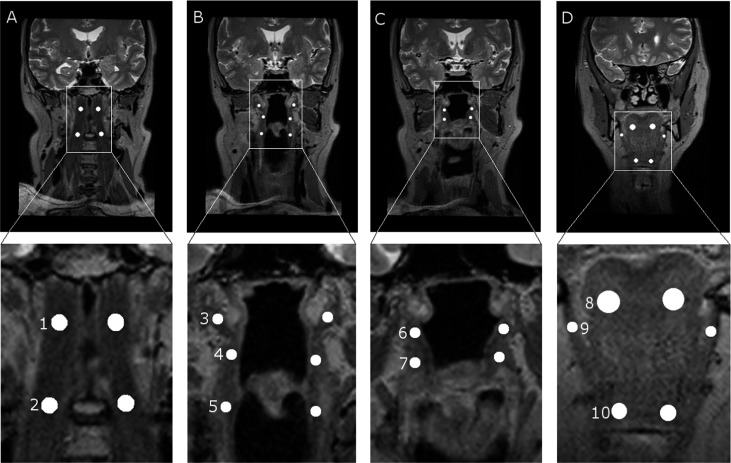
Figure 1.
Region of interest (ROI) locations in MR images. The upper row represents the original images, and the lower row represents the magnifications of ROI locations. (a) ROIs in longus capitis: 1, superior; and 2, inferior. (b) ROIs in pharyngeal muscles: 3, superior; 4, middle; and 5, inferior. (c) ROIs in palatal muscles: 6, tensor veli palatini; and 7, levator veli palatini. (d) ROIs in hyoid muscles: 8, genioglossus; 9, mylohyoideus; and 10, geniohyoideus.

