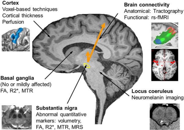
Figure 1.
Overview of MRI methods used to study PD. Cortex: changes were detected using voxel-based techniques, cortical thickness measurements and perfusion imaging. Brain connectivity was investigated using resting-state functional MRI (rs-fMRI) for functional connectivity and tractography for structural connectivity. Substantia nigra (yellow contour): changes were detected using diffusion imaging (reduced fractional anisotropy - FA), relaxometry (increased R2* indicating increased iron load and more recently susceptibility-weighted imaging), magnetization transfer ratio (MTR reduced) and spectroscopy. Basal ganglia: studies showed no or mild changes in FA, R2* or MTR. Locus coeruleus area (white arroxw head): reduced signal intensity was detected using neuromelanin imaging.

