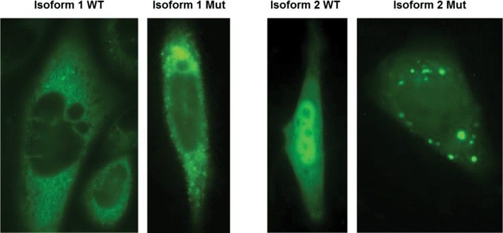
Figure 6.
Subcellular localization of METTL23-GFP fusion proteins for WT isoform 1, WT isoform 2 and the corresponding 5 bp-deletion mutant proteins are shown in CHO cells after transient transfection. The WT of isoform 1 is predominantly located in the ER and to a lower extent in the nucleus. The corresponding mutant protein shows a similar distribution but additionally forms highly fluorescent protein aggregates. The WT protein of isoform 2 appears to be located in comparable concentrations in the nucleus and in the cytoplasm, whereas the corresponding mutant protein located in the cytoplasm forms numerous concentric aggregates. Green fluorescent signal could also be observed in the nucleus.

