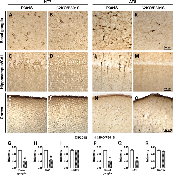
Figure 3.
Genetic suppression of β2ARs ameliorates neuropathology in hippocampus and basal ganglia of P301S mice. Photomicrographs of three commonly affected brain regions from P301S and β2KO/P301S mice. Sections were stained with the HT7 antibody, which recognizes total human tau, and AT8 antibody, which recognizes tau phosphorylated at Ser202/Thr205. (A and B) Representative basal ganglia sections demonstrate markedly reduced HT7 immunoreactivity in β2KO/P301S mice compared with P301S mice. (C and D) Representative sections of the hippocampal CA1 region. Effect similar to that in basal ganglia is observed with decreased HT7 immunoreactivity in β2KO/P301S mice. (E and F) Representative sections of neocortex demonstrates no difference in HT7 immunoreactivity between experimental and control mice. (G–I) The HT7 immunoreactivity was quantified using Image J (n = 6). The values in the y-axes are shown relative to P301S mice. (J and K) Representative basal ganglia sections demonstrate markedly reduced AT8 immunoreactivity in β2KO/P301S mice compared with P301S mice. (L and M) Representative sections of the hippocampal CA1 region. Effect similar to that in basal ganglia is observed with decreased AT8 immunoreactivity in β2KO/P301S mice. (N and O) Representative sections of neocortex demonstrate no difference in HT7 immunoreactivity between experimental and control mice. (P–R) The HT7 immunoreactivity was quantified using Image J (n = 6). The values in the y-axes are shown relative to P301S mice. Data are presented as means ± SEM and analyzed by Student's t-test.

