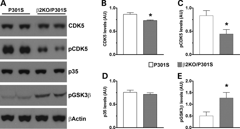
Figure 5.
CDK5 and GSK3β activities are reduced in β2KO/P301S mice. (A) Western blots of proteins extracted from the brains of P301S and β2KO/P301S mice. (B–E) Quantitative analyses of blots demonstrate that the levels of total CDK5 (B) and CDK5 phosphorylated at Tyr15 (C) are significantly higher in P301S mice compared with β2KO/P301S mice. However, the levels of p35 (D) remain unchanged. We also found that the levels of GSK3β phosphorylated at Ser9 (E) were significantly lower in the P301S mice compared with β2KO/P301S mice. Quantifications of the western blots were performed by normalizing the protein of interest to β-actin, which was used as a loading control. n = 6/genotype. Data are presented as means ± SEM and analyzed by Student's t-test. * indicates P < 0.05.

