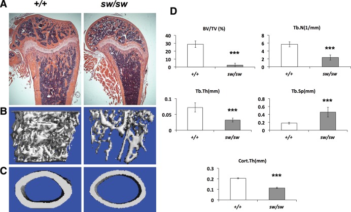
Figure 2.
The Wnt1sw/sw mice exhibit severe osteopenia. (A) H&E stained longitudinal sections of femurs of wild-type (+/+) and Wnt1sw/sw (sw/sw) mice at 6 weeks of age. μCT reconstruction image of trabecular bone (B) and cortical bone (C) at 6 weeks of age. (D) μCT analysis shows decreased bone volume per total volume (BV/TV), decreased trabecular number (Tb.N) and trabecular thickness (Tb. Th), increased trabecular separation (Tb. Sp), and decreased cortical bone thickness (Cort.Th) when compared with wild type (male mice, n = 6 per group, ***P < 0.001).

