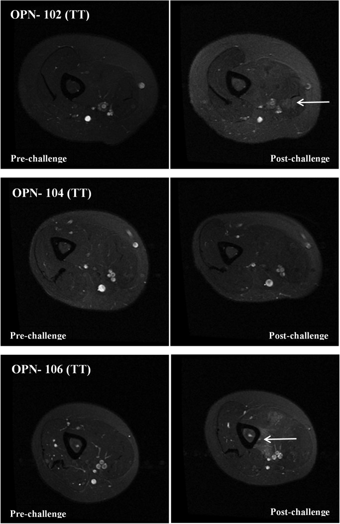
Figure 4.
T2-weighted MRI images of young adult female volunteers having TT OPN genotype. Shown are MRI T2-weighted images of the non-dominant upper arm prior to eccentric challenge (pre-challenge) and 4 days after eccentric challenge (post-challenge). Subjects OPN-102 and OPN-106 show areas with an increase of T2 signal in the biceps in the post-challenge images consistent with increased water content (swelling) (white arrows). Areas of hyper-intensity were not evident in OPN-104.

