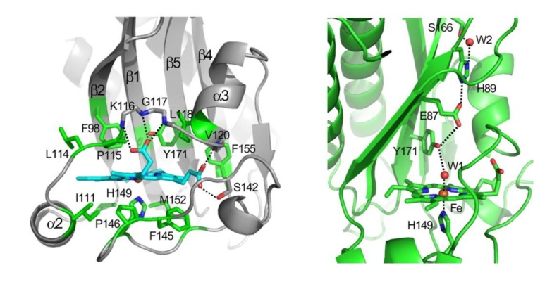
Figure 2.
Crystal structure of the ferric DosS GAF-A domain showing the heme surrounded by the hydrophobic residues in a ligand binding pocket. The heme iron is coordinated to His149 to the proximal side and the distal water molecule is hydrogen bonded to the Tyr171 residue, which interacts with the His89 via Glu87 [35]. Reprinted with permission. © 2008 The American Society for Biochemistry and Molecular Biology.

