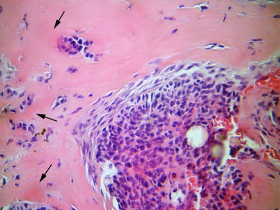
Fig. 5.

The cellular fibrous stroma and a nest of odontogenic epithelium can be seen. Foci of calcifications are in the lower right (hematoxylin and eosin stain, ×400). The arrows indicate the epithelial hyalinization areas (stronger pink coloration), suggestive of areas of induction
