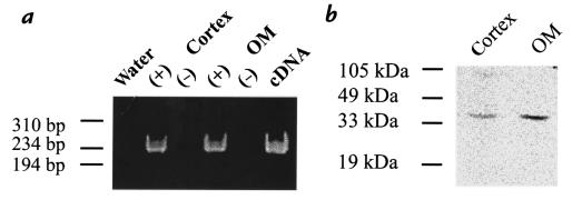
Figure 1.
Expression of HO-2 in rat kidney cortex and outer medulla (OM). (a) RT-PCR of rat kidney using primers specific for HO-2. An ethidium-stained 6% polyacrylamide gel is displayed; one-fifth of each PCR reaction was loaded per lane. A 230-bp fragment was amplified in rat cortex and outer medulla. Water and RT(–) controls are negative. (b) Western blotting of rat kidney, using an HO-2–specific antibody. RT, reverse transcriptase.

