
Fig. 3. Control immunostaining for K1, K10 and K17 in normal skin and basal cell carcinoma.
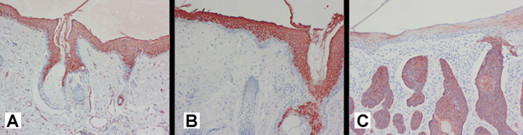
Intense immunostaining for (A) K1 and (B) K10 is present in the suprabasal keratinocytes of normal control skin (no immunostaining is detectable in the basal cell layer. 20×. (C) The dermal tumor aggregates of basal cell carcinoma show intense immunostaining for K17, while faint suprabasilar immunostaining is present in the overlying epidermis. 20×.