Fig. 1.
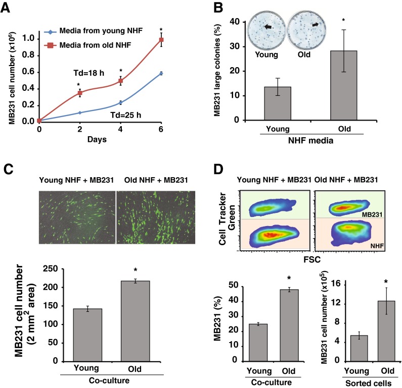
Aging quiescent fibroblast promotes breast cancer cell proliferation. a MB231 human breast epithelial cancer cells were cultured in conditioned media that were collected from young and old quiescent normal human fibroblasts (NHFs). Cell numbers were counted at 2 days interval, and cell doubling time was calculated from the exponential portion of the growth curve. Asterisks represent statistical significance between young and old cultures; n = 3, p < 0.05. b MB231 cells were plated at limited dilution for single cell colony assay. Cells were cultured in conditioned media for 14 days, fixed, and stained with Coomassie blue for visualization. Colonies with more than 150 cells were considered large colonies. Asterisks represent statistical significance between young and old cultures; n = 3, p < 0.05. c Upper panel: representative microscopy images of 4-day co-cultures of CellTracker green-labeled MB231 cells cultured on top of monolayers of young and old quiescent NHFs. Images of green fluorescence and bright field pictures were combined for the final image. Lower panel: NIH Image J software was used to count cells in 2 mm2 area. Asterisks represent statistical significance between young and old cultures; n = 5, p < 0.05. d Flow cytometry analysis of co-cultures. Upper panel: representative pseudo-color density plots of 5-day co-cultures of CellTracker green-labeled MB231 cells cultured on top of monolayers of young and old quiescent NHFs. Lower left panel: percentage of MB231 cells in co-cultures. Asterisks represent statistical significance between young and old cultures; n = 5, p < 0.05. Lower right panel: MB231 cells were sorted from co-cultures of NHFs by flow cytometry and cultured in regular media for 3 days prior to counting cells. Asterisks represent statistical significance between young and old cultures; n = 3, p < 0.05

