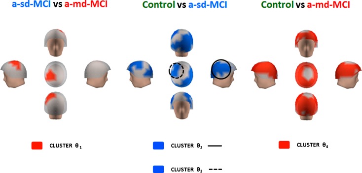
Fig. 5.
Differences within the theta band range. Red color indicates that the a-md-MCI group exhibited more relative power in left centro-parietal regions than a-sd-MCI group (cluster θ 1) and in practically the whole head when compared with control group (cluster θ 4). The a-sd-MCI exhibited a significant power increase compared with the control group in two clusters of sensors, represented in blue color, which involved the right lateral fronto-temporo-parieto-occipital region (cluster θ 2) and the left fronto-central location (cluster θ 3)

