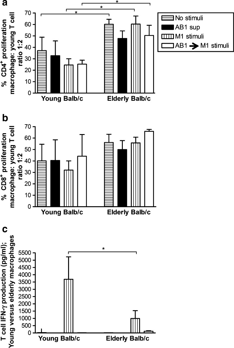
Fig. 2.
Tumor-exposed macrophages from elderly mice show impaired ability to stimulate T cell production of IFN-γ. Peritoneal macrophages from young or elderly Balb/c mice were cultured overnight with AB1 tumor cell-conditioned media (AB1 sup) then activated with M1 stimuli (LPS/IFN-γ) for another 24 h (AB1 sup → M1 stimulus). Controls included no stimuli, AB1 sup only, and M1 stimuli only. CFSE-labelled young C57BL/6J-derived splenic lymphocytes were added to macrophages at varying ratios and 5 days later cells stained for CD4 and CD8, and the percentage of proliferating CFSE+CD4+ T cells (a) and CFSE+CD8+ T cells calculated (b). MLR supernatants were assayed for T cell-derived IFN-γ by CBA; calculated by subtracting IFN-γ secreted by macrophages (c). Data from two to four experiments is shown at the macrophage: T cell ratio of 1:2 as mean ± SEM. *p < 0.05

