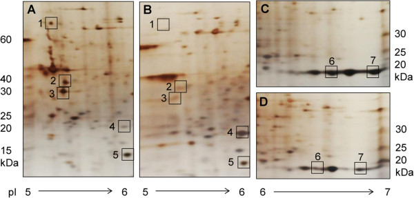
Figure 3.

Comparative 2D gel electrophoresis of highly (A and C) and weakly (B and D) pathogenic N. fowleri. The proteomes of trophozoites were separated via 2D gel electrophoresis, and differentially expressed proteins (squares) were excised and identified via nano-liquid chromatography tandem mass spectrometry (nano-LC MS/MS). In the figure, only enlarged images from gel segments with differential spot patterns are shown. The numbers correspond to the identified proteins listed in Table 3. Spot 5 was used as a control, representing a protein (cofilin) with equivalent expression in highly and weakly pathogenic trophozoites.
