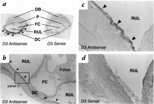
Figure 6.
In situ hybridization using the D3 antisense or sense probe on sections of a uterine implantation site on day E15. (a) Composite autoradiograph of adjacent longitudinal sections hybridized with the probes indicated. Sections are oriented so that the mesometrial surface of the uterus is toward the top. Specific signal is observed along the wall of the recanalized uterine lumen (arrowheads). (b) Low-powered and (c) higher-powered photomicrographs showing the antimesometrial portion of the section in a probed with the D3 antisense probe. Silver grains are noted over the epithelial cells lining the outer wall of the recanalized uterine lumen. In contrast, no specific signal is observed in the section hybridized with the D3 sense probe (d). DB, decidual basalis; DC, decidual capsularis; FC, fetal cavity; P, placenta; RUL, recanalized uterine lumen.

