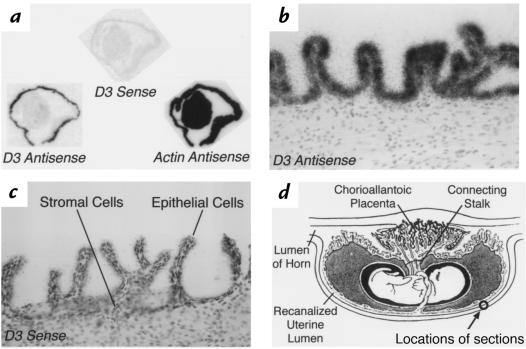
Figure 7.
In situ hybridization using D3 antisense, sense, or actin antisense probes on sections of an isolated pregnant uterus from E19. The specimen was harvested (as described in Methods) so that that the epithelial cells lining the recanalized uterine lumen lie on the outside of the specimen. (a) Composite autoradiograph of adjacent sections hybridized with the probes indicated. Specific signal with the D3 antisense probe is observed over the outer edges of the specimen. (b) High-powered light-field photomicrograph of a section hybridized with the D3 antisense probe showing intense signal over the epithelial cells. (c) Hybridization of an adjacent section with the D3 sense probe results in no specific signal. (d) Diagram of a longitudinal section through the fetal cavity and uterus of a late-stage rodent pregnancy, illustrating the approximate locations of the sections in this figure (reproduced, with permission, from ref. 74).

