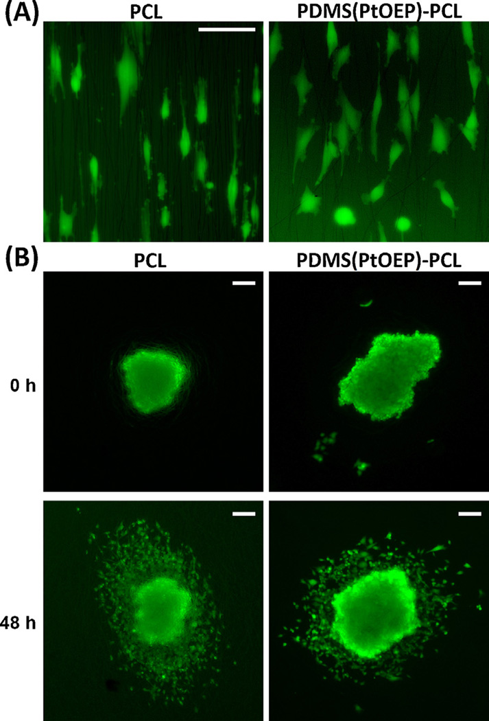
Fig. 6.
Cell morphology and dispersion on PCL nanofibers versus PDMS(PtOEP)-PCL nanofibers. (A) U251 cells show similar morphology and stretching over PCL and PDMS(PtOEP)-PCL fibers (bar: 50 µm). (B) Tumorspheres of U251 cells imaged immediately after deposition on randomly-aligned nanofibers and 48 hours later. Note the radial pattern of cell dispersion, which is similar in both types of fibers (bars: 100 µm).

