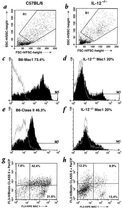
Figure 3.
Analysis of macrophage activation phenotypes in C57BL/6 (a, c, e, and g) vs. IL-12–/– mice (b, d, f, and h). Total lung mononuclear cells were isolated from two C57BL/6 and four IL-12–/– mice 27 days after infection, immunostained, and analyzed for Mac 1 and MHC class II expression by FACS. Gating was set up on macrophage-rich cell populations (a and b), and analysis was carried out to identify cells positive for Mac 1 (c and d), MHC class II (e and f), or both (g and h).

