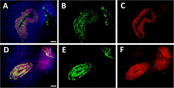
Figure 3.

Cldn6 was co-localized with TTF-1 at E12.5 (A-C) and E13.5 (D-F). Merged images are shown (A and D) that include Cldn6 immunofluorescence (B and F), TTF-1 immunofluorescence (C and F) and DAPI staining for cellular perspective. No immunoreactivity was observed in lung sections incubated without primary antibodies (not shown) and all images are at 40X original magnification. Scale bars represent 20 μm.
