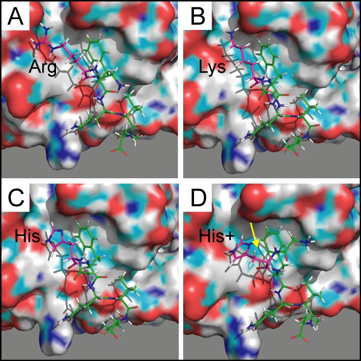
Figure 8. PyMol rendering of peptides and OSR1/CCT binding pocket.
A. GRFQVT wild-type peptide with arginine side chains extending towards negative charges within the pocket. B. GKFQVT mutant peptide with shorter side chain of lysine. C & D. GHFQVT mutant peptides are shown with or without protonation of the imidazole ring. Proton is highlighted with yellow arrow. The CCT/PF2 domains are rendered in surface mode, whereas the peptides are rendered in stick mode. The surface drawing of the domains highlights negative (red), positive (blue), and polar (green) moieties.

