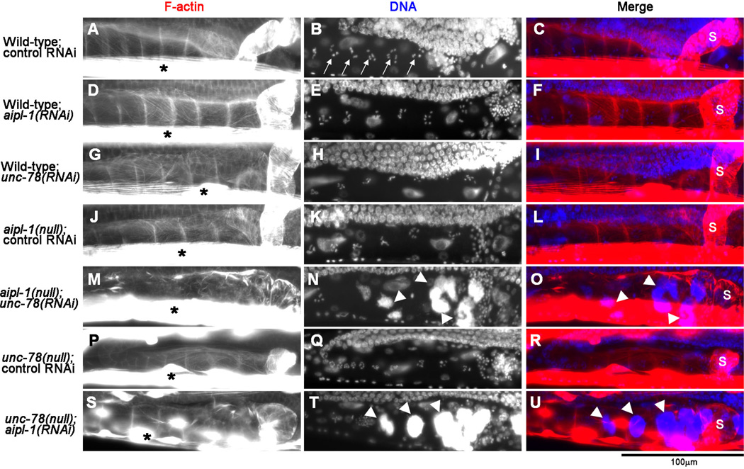
Fig. 1. Depletion of the two AIP1 isoforms causes accumulation of endomitotic oocytes in the ovary.
Adult worms with genotypes and RNAi treatments as indicated on the left were fixed and stained with tetramethylrhodamine-phalloidin for F-actin (left column) and DAPI for DNA (middle column), and regions of the ovary and the spermatheca are shown. The spermatheca is on the right side of each micrograph, and its location indicated by “s” in merged images on the right column (red: F-actin; blue: DNA). Arrows in B indicate condensed chromosomes in normal oocytes. Asterisks in the left column show intense staining of F-actin in the body wall muscle. Arrowheads in N and T indicate abnormal accumulation of DNA in endomitotic oocytes. Bar, 100 µm.

