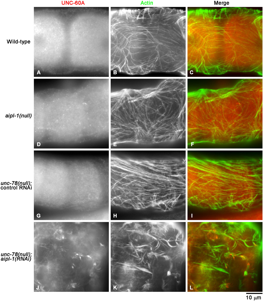
Fig. 6. UNC-60A (ADF/cofilin) is mislocalized to actin aggregates in AIP1-depleted myoepithelial sheath.
Gonads from worms with genotypes and RNAi treatments as indicated on the left were dissected out and stained with anti-UNC-60A antibody (left column) and anti-actin antibody (middle column), and the regions of the myoepithelial sheath are shown. Merged images are shown in the right column (red: UNC-60A; green: actin). UNC-60A was expressed in both oocytes and the myoepithelial sheath. Although the focuses were adjusted to the levels of the myoepithelial sheath, staining of oocytes were also visible. Bar, 10 µm.

