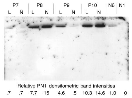
Figure 4.
Media (conditioned for 48 h) from four scleroderma fibroblast lines, P7–P10 (lesional and nonlesional; L and N, respectively), and two normal dermal fibroblast lines, N6 and N1, were used for Western blotting. One of the two normal lines showed no detectable PN1 (N1). The other normal line was used as a baseline, and its value for secreted PN1 was arbitrarily set to 1.0. Protein concentrations were determined and equal amounts loaded in each lane. The values for the PN1 levels secreted by the scleroderma lines are shown beneath each lane.

