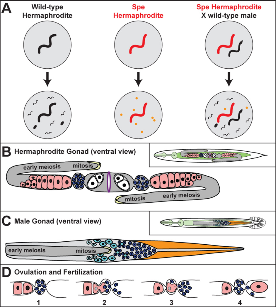
Figure 1. Reproduction in C. elegans.
A. Examples of self and cross fertilization in wild-type and mutant C. elegans. The male has an angular tail. Eggs are black ovals, and unfertilized oocytes are orange circles. B. The hermaphrodite gonad. Oocytes are pink, sperm blue, eggs white, the distal tip cells yellow and the vulva purple. The inset shows the position of the gonad in the body. Cells in mitosis or early meiosis are part of a syncytium. C. The male gonad. Cells in mitosis or early meiosis are part of a syncytium. Primary spermatocytes are light blue cells, residual bodies are light blue circles, and spermatids are dark blue circles. Distal tip cells are yellow, and the vas deferens orange. The inset shows its position in the male body. D. Ovulation and fertilization. (1) The animal is preparing to ovulate a mature oocyte (located on the right). (2) The mature oocyte nucleus undergoes nuclear envelope breakdown. During ovulation, the oocyte enters the spermatheca and is fertilized by a sperm. (3) The resulting zygote pushes many other sperm into the uterus. (4) The zygote commences embryogenesis as it passes into the uterus and sperm crawl back into the spermatheca for the next cycle.

