FIGURE 4.
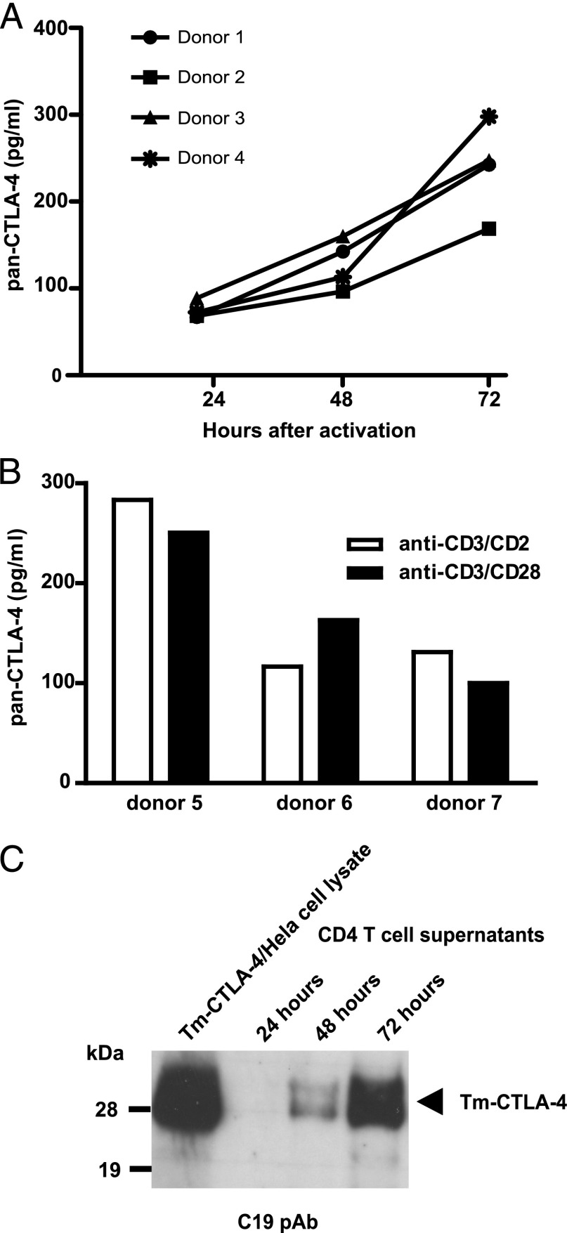
Immunoassay and Western blot analysis of Tm-CTLA-4 present in culture supernatants from activated CD4+ T cells. (A) Culture supernatants from 3 × 10⁶ cells/ml CD4+ T cells activated with anti-CD3/CD2 mAbs in vitro from four donors cultured on different days were harvested at the indicated time points and analyzed using the pan–CTLA-4 immunoassay. (B) Analyses of supernatants of CD4+ T cells (1 × 10⁶ cells/ml) of three donors cultured on different days stimulated in vitro for 72 h with either anti-CD3/CD2 mAbs or anti-CD3/CD28 microbeads. (C) Release of Tm-CTLA-4 in a time-dependent manner in culture supernatants of activated CD4+ T cells was confirmed by Western blot analysis (reducing gel) using Tm-CTLA-4 C terminus C19 polyclonal Ab. Western blot gel is representative of at least four independent experiments.

