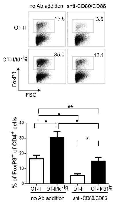
Figure 5. In vitro Treg differentiation on thymic dendritic cells.
CD4+CD25−CD69+ thymocytes sorted from OT-II or OT-II/Id1tg mice were co-cultured with purified thymic DC in the presence of 40 μg/ml ovalbumin, 10 ng/mL recombinant IL-2 and 1ng/ml IL-7 for 4 days. Representative FACS plots of FoxP3 expression in CD4 single positive T cells are shown on the top. Data from five independent experiments are presented as mean with SD at the bottom. **p<0.01; *p<0.05; ns, not significant.

