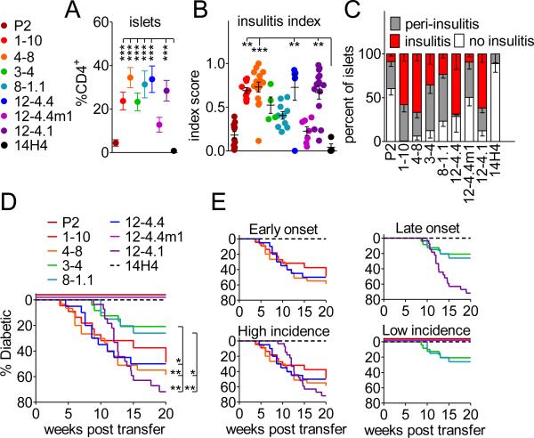
FIGURE 2. High and low affinity TCRs are able to accumulate in the pancreas and induce spontaneous diabetes development.
(A) Frequencies of insulin specific T cells in the infiltrated pancreatic islets were obtained from analysis of TCR retrogenic mice 7-8 weeks post bone marrow transfer. An average of at least 10 mice from 4 separate experiments is shown. Error bars represent SEM.
(B and C) Histological assessment of insulitis in insulin TCR retrogenic mice at 8-9 weeks post bone marrow transfer.
(D) TCR retrogenic mice were monitored for spontaneous diabetes development (n ≥ 20 mice per group).

